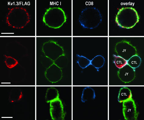
Fig. 3.
Enrichment of Kv1.3/FLAG in or around the IS. Distribution of Kv1.3/FLAG (red: anti-FLAG followed by Alexa Fluor 546-RAMIG), MHC class I (green: X-FITC-W6/32), and CD8 (Alexa Fluor 647-anti-CD8) molecules in a lone CTL (Top) and CTL-JY target cell conjugates (Middle and Bottom). The thickness of the optical slice was 1.1 μm. In lone CTLs, the channels are distributed in small patches throughout the membrane (Top). On CTL-target interaction Kv1.3 channels are redistributed and enriched in the IS in the majority of CTLs (Middle) or surround the synapse like a ring (Bottom). (Scale bar, 5 μm.)

