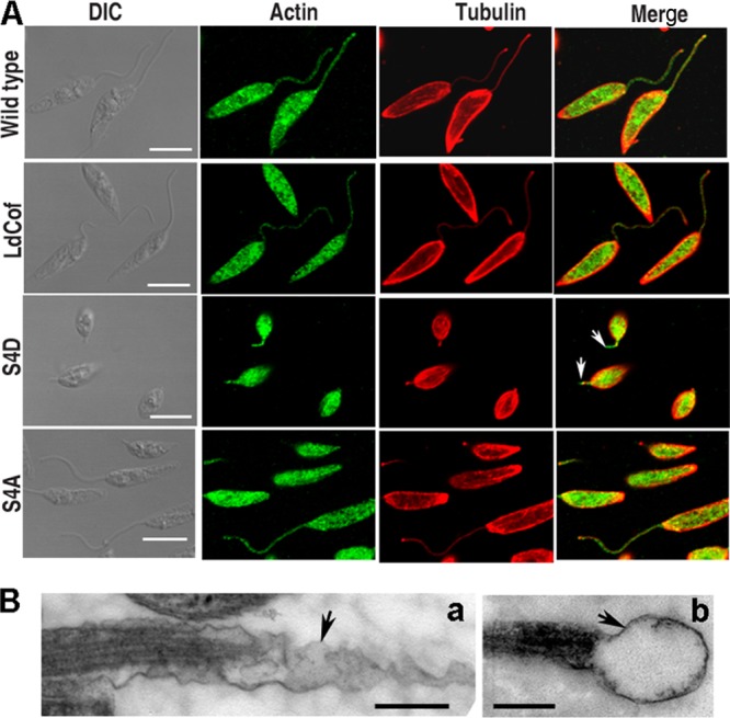
Fig 8.

(A) Analysis of actin distribution and length of flagellar axoneme in LdCof, S4D, and S4A protein-overexpressing cells. In wild-type and overexpressing cells, actin and tubulin were stained with polyclonal rabbit anti-Leishmania actin antibodies and polyclonal mouse anti-α,β-tubulin antibodies, respectively. Actin distributions in S4D and S4A protein-overexpressing cells were similar to that in wild-type cells. A flagellar sleeve (membrane extension without axoneme [16]) was clearly visible beyond the length of the axoneme (stained with antitubulin antibodies and marked by an arrow) in the flagella of S4D protein-overexpressing cells. Bar, 5 μm. (B) Ultrastructure of flagellar sleeve (arrows) in S4D-overexpressing Leishmania cells by transmission electron microscopy. Elongated (a) and bulbous (b) membrane extensions beyond the axoneme extremity are seen. Bar, 200 nm.
