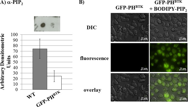
Fig 7.
GFP-PHBTK-expressing cells have altered PIP2 levels, and can be loaded with PIP2. (A) Phosphoinositides were extracted from whole-cell lysates, and PIP2 levels were measured using dot blots with antibodies specific to PIP2. Levels were analyzed and assigned a value of arbitrary densitometric units. PIP2 levels were lower in GFP-PHBTK-expressing cells than in wild-type cells. (B) PIP2 loading in GFP-PHBTK-expressing cells was confirmed using a BODIPY-labeled PIP2. Both differential interference contrast (DIC) and fluorescence images are shown. Scale bars represent 25 μm.

