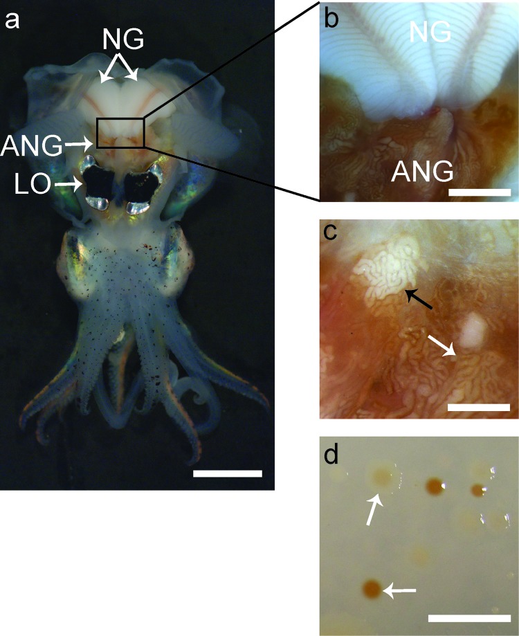
Fig 1.
Anatomy of a female Euprymna scolopes and morphology of ANG isolates. (a) Ventral dissection of E. scolopes, showing the accessory nidamental gland (ANG) located posterior to the light organ (LO) and in close proximity to the nidamental gland (NG). (b) Pigmented ducts in the NG converge at the ANG. (c) Magnification of the ANG reveals convoluted tubules, most of which are dark orange in pigmentation (white arrow), but others appear white (black arrow) or yellow (not shown). (d) Culturing of ANG symbionts results in many colonies with pigmentation similar to that of the ANG tubules (white arrows). Bars, 1 cm for panels a and d, 2.5 mm for panel b, and 1 mm for panel c.

