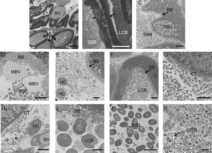
Fig 2.
Light microscopy and TEM of fixed sections from the E. scolopes ANG. (A) Cross-section of ANG tissue, showing many tubules interspersed between blood vessels (BV). Most tubules contained dense bacterial populations (T); however, some were empty (ET). (B) Closer inspection revealed that tubules have two distinct morphotypes, comprising a large coccoid bacterium (LCB) and a smaller bacillus-shaped bacterium (SBB) that were observed in tubules with differing epithelial morphologies, either a vacuole-rich epithelium (arrowhead) or dense epithelium (black arrow). (C) Tubule with brush border (BB) dominated by SBB with an epithelial morphology suggesting secretory cells (SEC). (D) SBB inhabiting tubules with a thick microvillar brush border (BB) and membrane-bound vesicles (MBV). (E) Hemocytes (HC) in the lumen of a tubule with a population of bacteria. (F) Tubule dominated by LCB. (G) Bacteria were also seen in the connective tissue (CT) outside the tubules; note the absence of a brush border. (H) Hemocytes were also seen with bacteria among the connective tissue. (I) Closer inspection of the LCB revealed many storage granules. (J and K) One SBB morphotype showed a nucleoid structure, while the other (K) showed many polyhydroxybutyrate-like granules (black arrow). Bars, 30 μm (A and B), 5 μm (C to H), and 1 μm (I to K).

