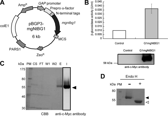
Fig 1.
Cloning, expression, purification, and deglycosylation assay. (A) Expression plasmid. (B) Production of G1mgNtBG1 was analyzed by an enzyme activity assay (upper graphic) and a Western blot assay with anti-c-Myc antibody (bottom panel) and compared with that of the control. (C) Purification was performed by Ni2+-NTA chromatography. Fractions were resolved by SDS-PAGE, and the gels were either stained with CBB or transferred and immunoblotted with anti-c-Myc antibody. PM, protein marker; CS, culture supernatant; FT, flowthrough; W1 and W2, wash fractions 1 and 2; E, elution fraction; I, immunoblotting. The arrowhead indicates the G1mgNtBG1 band. (D) G1mgNtBG1 was treated (+) or not treated (−) with Endo H, separated by SDS-PAGE, and stained with CBB. The reduction in size from 60 kDa (closed arrowhead) to 56 kDa (open arrowhead) indicates the presence of the posttranslational modification by N-glycosylation.

