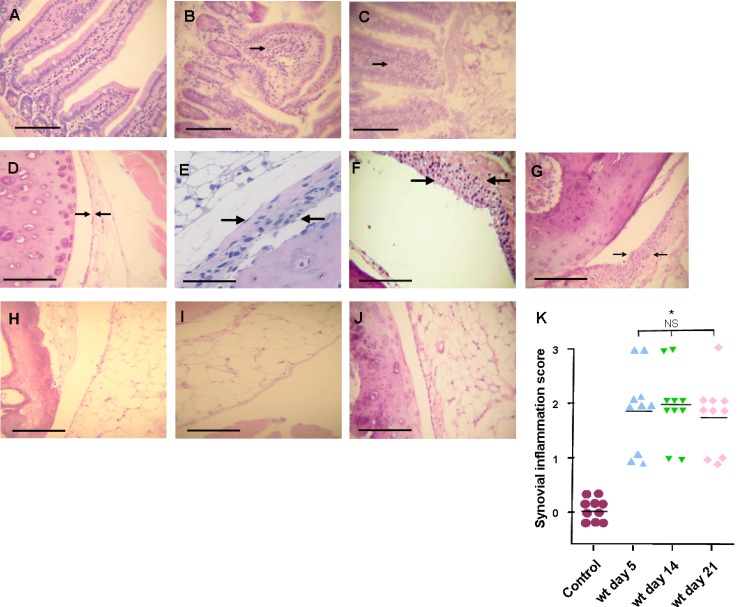
Fig 1.
Histological changes in large intestine and joints after S. Enteritidis enterocolitis. Histology studies after intragastric inoculation with 3 × 10³ to 4 × 10³ CFU of the wild type (wt) or 107 CFU of the invG mutant of S. Enteritidis (ΔinvG). (A, B, and C) Large intestine. (A) Control animals: normal display of intestinal mucosa; (B) Enterocolitis in wt infected mice, note the loss of normal villi display and the infiltration of mononuclear cells (arrow); (C) enterocolitis in mice inoculated with the ΔinvG strain, changes are similar to those observed in the wt group. HE stain. Bar: 100 μm. (D to J) Joints. (D) Control animals: normal synovial capsule (arrows). (E, F, and G) Mice infected with the wt strain: moderate hyperplasia, with 3 to 5 layers of synoviocytes (arrows) at days 5, 14, and 21, respectively. (H, I, and J) Mice infected with the ΔinvG strain: no differences were found with respect to control mice (D) at any time point studied (days 5, 14, and 21, respectively). HE stain. Bar, 1 mm (A, C, D, E, F and G) or 100 μm (B). (K) Synovial inflammation scores. *, significant differences (P < 0.01) between wt-infected mice and control group mice were found at all time points assessed. NS, no significant differences among wt-infected mice. Data were collected from three independent experiments.

