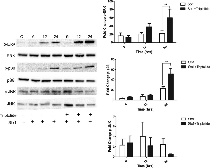
Fig 5.
MAPK phosphorylation in macrophage-like THP-1 cells treated with Stx1 and triptolide. Cells were stimulated with Stx1 (400 ng/ml) in the presence or absence of 0.05 μM triptolide for various time points. Whole-cell lysates (70 μg) were subjected to SDS–4 to 15% PAGE and probed with polyclonal antibodies against phospho-ERK, phospho-JNK, phospho-p38, ERK, JNK, and p38 MAPKs. C, untreated controls. The blots shown are representative of three to four independent experiments. The bar graphs represent mean fold change ± SEM calculated from densitometric scanning of three to four individual experiments for phospho-ERK (p-ERK), phospho-JNK (p-JNK), and phospho-p38 (p-p38) levels normalized to total MAPKs. Statistical significance was calculated using one-way ANOVA (**, P <0.01).

