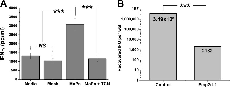
Fig 2.
PmpG1.1 recognition of infected epithelial cells and termination of C. muridarum replication within them. (A) PmpG1.1 cells were cocultured 1:1 with C57epi.1 cells that were mock infected or 18 h postinfection with 5 IFU per cell (MoPn), without and with tetracycline (TCN) cotreatment; IFN-γ in 24-h culture supernatants was quantified by ELISA. (B) C57epi.1 monolayers, pretreated for 12 h with 10 ng/ml IFN-γ, were infected with C. muridarum (3 IFU per cell). Four hours later, the monolayers were washed and then cocultured with PmpG1.1 T cells at an effector-to-target ratio of 0.75:1. The contents of the wells were harvested 32 h postinfection, and the recovered IFU were enumerated on McCoy monolayers. The mean number of IFU recovered for each condition is shown within the bars. Panels A and B represent single experiments done in quadruplicate. ***, P < 0.0005; NS, not statistically significant. The error bars indicate SD.

