Fig. 3.
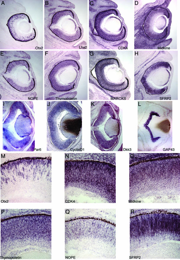
Expression of genes identified as progenitor cell-enriched. In situ hybridization studies of the expression of a subset of the core set of retinal progenitor cell-enriched transcripts. All sections are from the E14.5 mouse retina. (A–L) Low-power views of expression of this subset of genes. Gene identifiers are as shown on each panel. Retinal progenitor cells occupy over three-quarters of the radial thickness of the neural retina at this stage with the innermost cells being ganglion cells, as shown by the neuron-specific GAP-43 staining in L. Note that although most genes are expressed in progenitor cells (for example, in A, B, and D), some genes are expressed in both progenitor cells and neurons (for example, in C and G). (M–R) High-power views of the progenitor cell-specific expression of a selection of genes, as labeled in each panel. Note the lack of expression of five of the six genes on the inner (vitreal) side of the retina populated by ganglion and amacrine cells, with CDK4 expressed at a low level within this region. Examples are also shown of genes with heterogeneous expression within progenitor cells, including Otx2 (M) and NOPE (Q).

