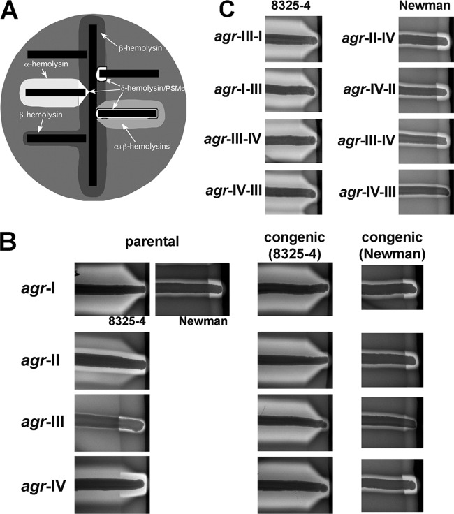
Fig 3.
Analysis of hemolytic activity of S. aureus strains on sheep's blood agar (SBA). (A) Schematic of hemolytic activities on SBA. Tested bacteria (horizontal black bars) are streaked at a right angle to RN4220 (vertical black bar), a β-hemolysin producer, and the plate is incubated overnight. β-Hemolysin forms a turbid zone of hemolysis; β is synergistic with δ-hemolysin and other PSMs, producing an amplified zone of clearing where they intersect; β inhibits α-hemolysin, producing the V-shaped zone of clearance where they intersect. (B, C) Hemolytic activities on SBA for parental and congenic strains with the indicated agr allele cross-streaked with RN4220. Images from each column are from the same plate. Parental strains: agr-II, RN6607; agr-III, RN3964; agr-IV, RN4850. The pattern seen with native agr-II is due to the production of β-hemolysin, as well as α and δ and/or other PSMs; Newman has a prophage in the β-hemolysin gene. The α-β mutual inhibition is seen only with Newman agr-III because of the low δ-hemolysin and/or other PSM production by group III. In panel C, the chimeras tested correspond to those tested for reporter activity in Fig. 2C and D.

