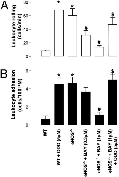
Fig. 3.
Leukocyte–endothelial cell interactions [rolling (A) and adhesion (B)] in mouse mesenteric postcapillary venules in vivo in WT and eNOS–/– mice in the absence and presence of BAY 41–2272 (0.3–1 μM) and ODQ (5 μM). *, P < 0.05, significantly greater than WT animals; #, P < 0.05, significantly lower than eNOS–/– mice; $, P < 0.05, significantly greater than eNOS–/– mice with BAY 41–2272 alone; n > 5.

