Fig 7.
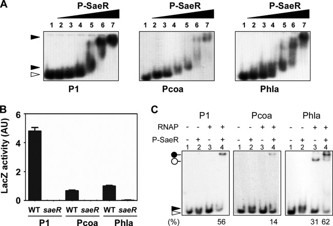
RNAP can bind to Phla without P-SaeR. (A) SaeR binding of the three target promoters. Promoters (2 ng) labeled with 32P were mixed with 3 μg/ml salmon sperm DNA and 0 μM, 0.25 μM, 0.5 μM, 1 μM, 2 μM, 4 μM, or 8 μM P-SaeR (lanes 1 to 7); incubated at room temperature for 15 min; and analyzed by 5% PAGE and autoradiography. The white arrowhead indicates free probes. (B) Dependence of the three sae target promoters on the SaeRS TCS. Promoter-lacZ fusion plasmids were inserted into WT or saeR mutant strain Newman, and then promoter activity was measured by lacZ expression. saeR, saeR mutant. Data are representative of results obtained from three independent experiments. Error bars represent standard deviations. (C) Binding of RNAP to the three target promoters. DNA probes were mixed with RNAP (0.7 μg) and/or P-SaeR (0.5 μM), incubated for 15 min at room temperature, and then analyzed by 5% PAGE and autoradiography. The white and black arrowheads indicate free and P-SaeR-bound probes, respectively. The white pinhead denotes the DNA probe-RNAP complex, and the black pinhead represents the DNA probe–P-SaeR–RNAP ternary complex. The percentage of DNA probe in the protein-DNA complex is shown at the bottom.

