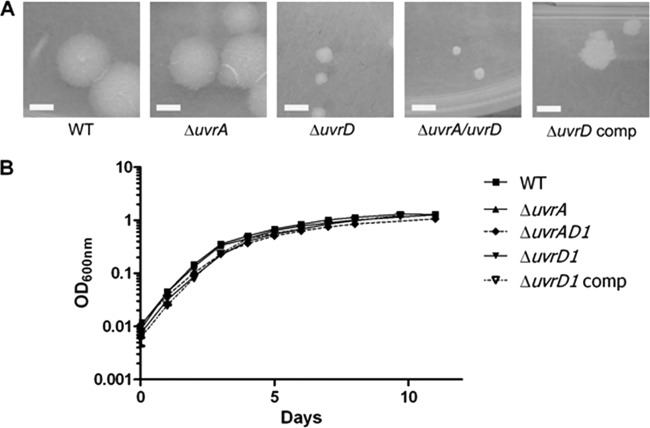
Fig 2.
In vitro growth characteristics of the uvrD1 and uvrA uvrD1 mutant strains. (A) The colony size and morphology of the uvrD1 and uvrA uvrD1 strains are compared with the wild type, the uvrD1 complement, and a uvrA mutant, demonstrating the reduced colony size caused by inactivation of uvrD1. In each case the white bar in the image represents 3 mm, and the images were taken after 3 weeks of growth at 37°C. (B) Growth of the strains in liquid culture was compared with that of the wild-type parental strain by measuring optical density.

