Figure 3.
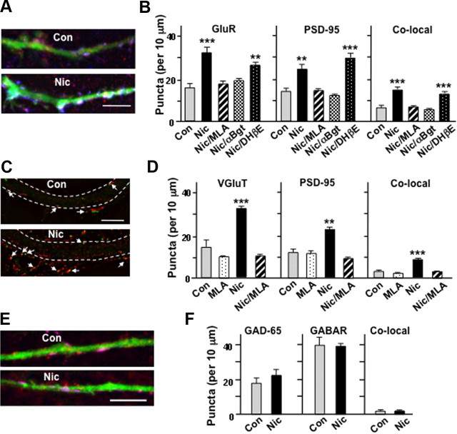
Nicotine in cell culture acts via α7-nAChRs to increase glutamatergic synaptic contacts. A, Control (Con) and nicotine-treated (Nic) cell cultures expressing GFP and surface stained for GluR1 receptors (red) and then intracellular PSD-95 (blue). Here and below, overlap of three colors appears as white. Scale bar, 10 μm. B, Quantification showing a significant increase in GluR1, PSD-95, and colocalized puncta in nicotine unless α7-nAChRs were blocked with either MLA or αBgt (n = 3–12 cultures, 5 cells/culture). C, Cultures immunostained for VGluT (red) and PSD-95 (green). The dashed white lines indicate the edges of a neurite; spines extending from the neurite are not demarcated. Scale bar, 5 μm. D, Quantification showing increased puncta and colocalization of VGluT and PSD-95 after nicotinic activation of α7-nAChRs. E, Cultures expressing GFP and immunostained first for surface α1-containing GABAA receptors (red) and then for intracellular GAD-65 (blue). Scale bar, 10 μm. F, Quantification showing no changes in the numbers of GAD-65 and GABAA receptor puncta (GABAR) or in their colocalization (n = 3–4 cultures; 5 cells/culture). Nicotinic stimulation appears to increase glutamatergic, but not GABAergic, synapses.

