
Figure 6.
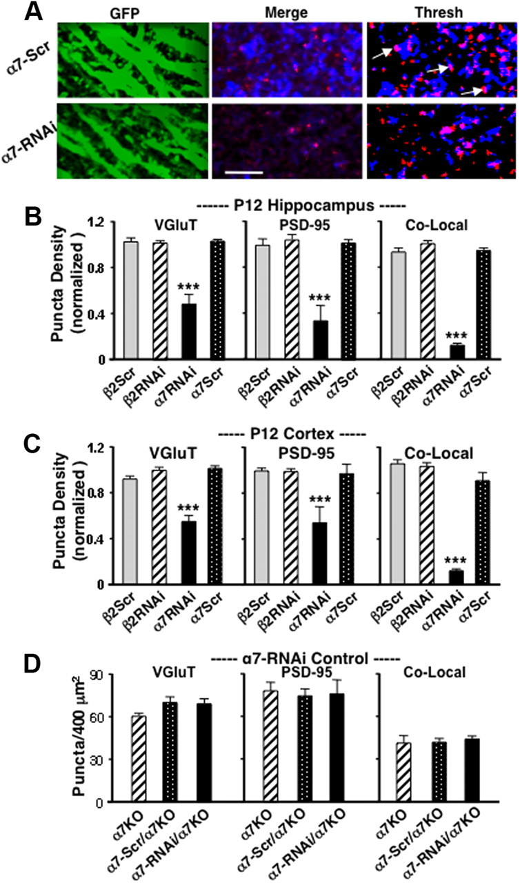
Relevance of local α7-nAChRs for glutamatergic synapse formation. A, Apical dendrite regions of P12 hippocampal CA1 expressing either lentiviral α7-Scr (upper) or α7-RNAi (lower) after intracranial injection in vivo (GFP) and immunostaining for VGluT (blue) and PSD-95 (red) shown together (Merge) and after thresholding (Thresh; arrows indicate examples of colocalization). Scale bar, 5 μm. B, Quantification of VGluT, PSD-95, and colocalized puncta/400 μm2 in P12 hippocampal CA1. C, Quantification in P12 visual cortex layer 5/6. Results are normalized to values obtained in adjacent regions lacking viral infection and demonstrate that α7-nAChRs within the local area are required for neurons to acquire WT levels of glutamatergic synapses. D, Quantification of VGluT, PSD-95, and colocalized puncta in α7KOs expressing the indicated constructs, showing that the α7-RNAi construct has no off-target effects as defined by changes induced in α7KOs (3 fields/animal; 4–8 animals/condition).