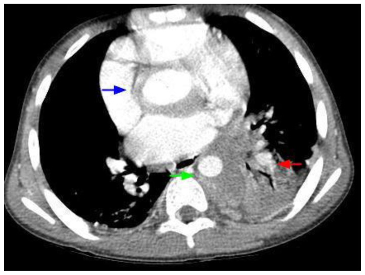
Figure 3.
12-year-old male with Rosai-Dorfman disease. Axial chest CT obtained following the intravenous administration of 100 ml of Omnipaque 350. The CT setting was 100 kVp with modulated mAs. The image reveals a mass encasing the aortic root (blue arrow). There is left lower lobe atelectasis (red arrow) secondary to compression of the left lower lobe bronchus. The mass is noted to encircle the descending aorta (green arrow) and compress the esophagus.

