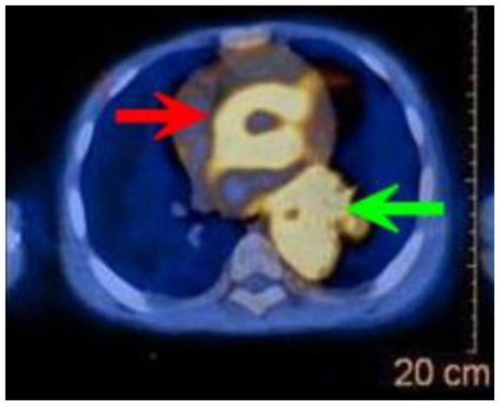
Figure 9.
12-year-old male with Rosai-Dorfman disease. Axial view of a PET/CT obtained following the intravenous administration of 8.7 mCi of F-18-FDG. The enhanced CT portion of the study was obtained following the intravenous administration of 100 ml of Omnipaque 350. The CT setting was 100 kVp with modulated mAs. There is intense hypermetabolism in the mass that surrounds the ascending and the descending aorta. The maximal SUV measured in the mass was 9.8, significantly higher than the blood pool activity.

