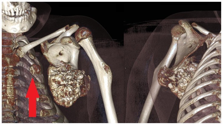
Figure 3.
A 6 year-old girl with a large scapular osteochondroma complicating congenital diaphyseal aclasia. Volume rendered images from the non-contrast CT demonstrate anterior and posterior views the lesion arising from the inferior angle of the scapula. Note also another osteochondroma arising from the 2nd anterior rib (arrow). (Protocol: 128 slice non-contrast CT, slice thickness 1mm, kVp 120, mAs 29).

