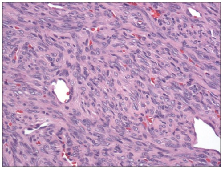
Figure 2.
A 57-year-old male with multifocal solitary fibrous tumors of the retroperitoneum and pancreas.
200× magnification of the solitary fibrous tumor using hematoxylin and eosin stain. The tumor displayed a classic “patternless” pattern of hyper- and hypocellular spindle cell areas, with prominent vasculature.

