Figure 3.
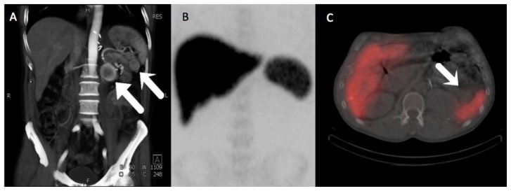
A 57-year-old male with multifocal solitary fibrous tumors of the retroperitoneum and pancreas.
A) Three dimensional volume-rendered MIP images from a CT angiographic study of the abdomen and pelvis show avid peripheral enhancement of the retroperitoneal mass, whereas the pancreatic tail mass has enhancement that is very similar to spleen. B) Anteroposterior MIP of the abdomen and pelvis from a subsequent Tc-99m sulfur colloid scan, showing normal uptake in the reticuloendothelial system. C) Fusion SPECT/CT image from the Tc-99m sulfur colloid scan shows no significant uptake of radiotracer within the pancreatic tail mass in distinction to the high activity seen within the adjacent spleen, proving the mass is not a splenule.
(Protocol: Splenic CT Protocol, 225 mAs, 120 kV, 0.75 mm slice thickness, 120 mL Omnipaque 350. Protocol: Anteroposterior MIP of the abdomen and pelvis from a subsequent Tc-99m sulfur colloid scan and Fusion SPECT/CT image from the Tc-99m sulfur colloid scan, 5.467 mCi Tc-99m sulfur colloid given IV, Imaging performed at 5 minutes, non-contrast, non-diagnostic CT performed for attenuation correction with 4.5 mm slice thickness.)

