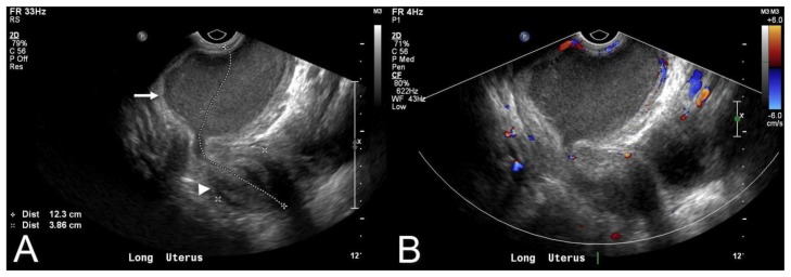
Figure 2.
17 year old female with OHVIRA and pyocolpos. Endovaginal longitudinal view of the uterus (8 MHz EV transducer, Phillips iU22) demonstrating an obstructed left hemivagina (arrow) with retained echogenic fluid. Uterus is also visualized in this image (arrowhead). Color Doppler demonstrates no flow in the collection.

