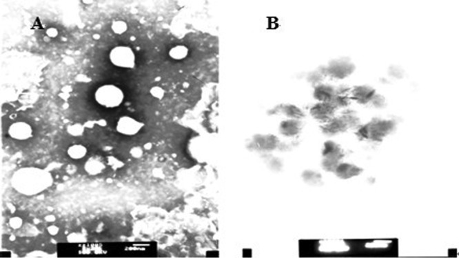
Fig 5.

Transmission electron microscopy of polybia-CP-treated LUVs. Transmission electron micrographs showing the morphological changes in liposomes composed of EYPE-EYPG (7:3) in the absence (A) and presence (B) of polybia-CP. The inset showed that polybia-CP could disrupt the integrity of liposome. Bar, 200 nm.
