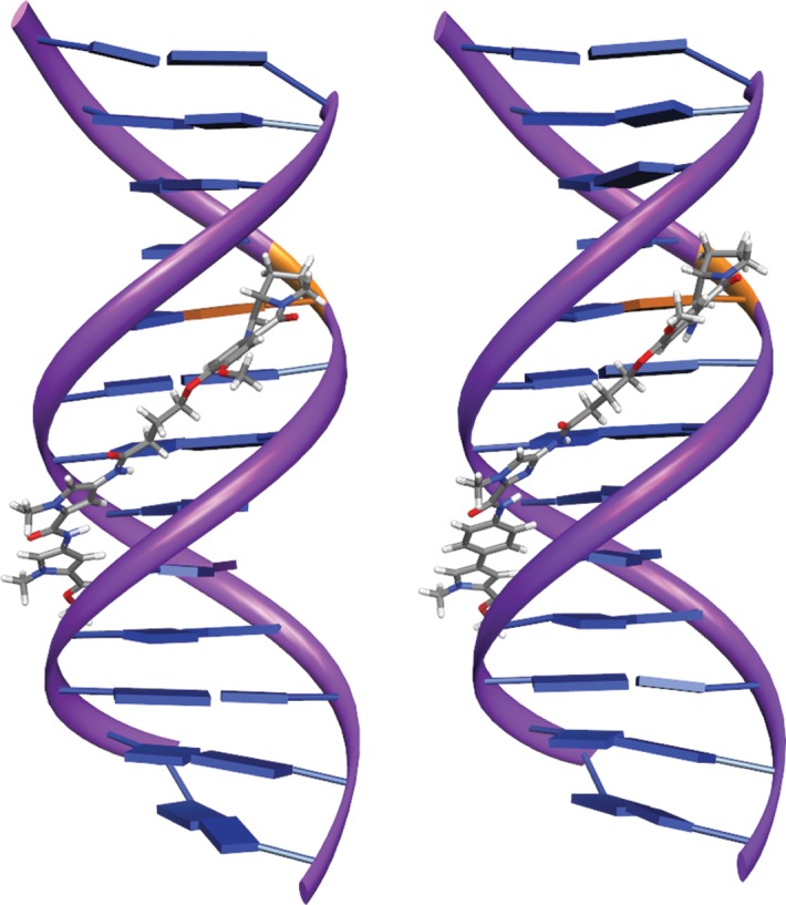
Figure 6.

In silico energy-minimized structures of the PBD binding target sequence TATAGAATCTATA and compound 24 Py-Py-PBD (left) and compound 36 MPB-Im-PBD (right). The covalent binding site is shown in orange. This figure appears in colour in the online version of JAC and in black and white in the print version of JAC.
