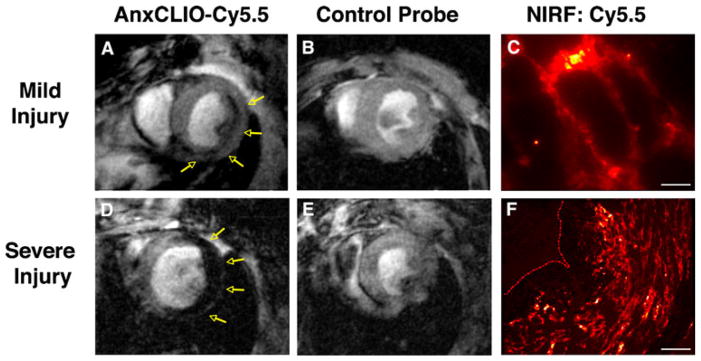
Fig. 1.
Molecular MRI of cardiomyocyte apoptosis with AnxCLIO-Cy5.5. a In mice with mild–moderate injury, the uptake of AnxCLIO-Cy5.5 is confined to the midmyocardium and b no uptake of the control probe is seen. d In mice with severe injury, the uptake of AnxCLIO-Cy5.5 is transmural and e minimal retention of the control probe is seen. MRI in this study was performed 4 h after ischemia–reperfusion injury, which is significantly earlier than previous studies with radiolabeled annexin. The fluorochrome on the agent allows the cellular distribution of AnxCLIO-Cy5.5 to be confirmed with fluorescence microscopy (high magnification in c, scale bar represents 20 m; low magnification in f, scale bar represents 80 m); reproduced with permission from [7]

