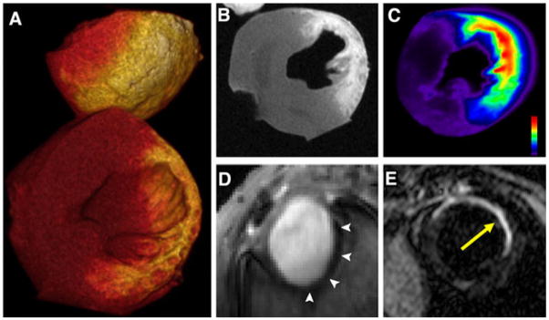
Fig. 3.
Molecular imaging of infarct evolution. a–c Loss of cardiomyocyte viability in acute infarction detected with delayed enhancement of Gd-DTPA-NBD in a heart excised 20 min after injection of the probe. Loss of cell membrane integrity expands the extracellular space leading to accumulation of the agent. a Volume rendered 3D MRI, b 2D short-axis reconstruction at midventricular level, and c fluorescent reflectance imaging. The accumulation of Gd-DTPA-NBD is well resolved with both MRI and fluorescence imaging. Reproduced with permission from [7]. d Angiogenesis in subacute healing infarcts: short-axis gradient-echo image of an infarcted mouse injected with integrin-sensing and Gd-loaded quantum dots (cNGR-pQDs). The r1/r2 ratio of the quantum dot differs significantly from small Gd chelates, producing negative enhancement (arrows); reproduced with permission from [27]. e Scar formation in chronic infarcts: MRI of myocardial fibrosis in mice with chronic infarcts injected with a collagen-targeted contrast agent (yellow arrow); reproduced with permission from [29]

