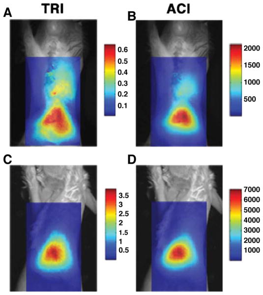
Fig. 5.
Planar fluorescence imaging of near-infrared fluorochromes. An infarcted mouse (a, b) and a sham-operated mouse (c, d) have been injected with CLIO-Cy5.5. The probe is taken up by macrophages infiltrating the healing infarct and can be imaged with fluorescence. The fluorescence images have been superimposed over white light images and two different postprocessing schemes have been used: TRI transillumination ratio image and ACI attenuation corrected image. While substantial hepatic uptake of CLIO-Cy5.5 is seen in both mice due to clearance of the probe in the liver, thoracic uptake of the agent is seen only in infarcted mice. Reproduced with permission from [10]

