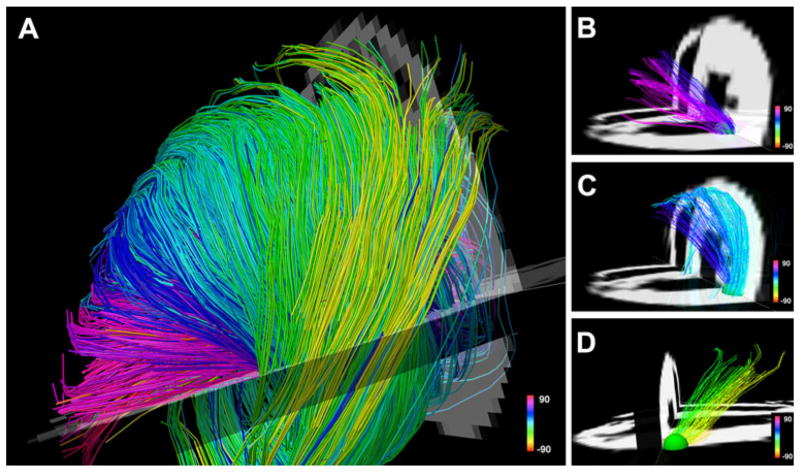
Fig. 8.
Myofiber architecture in a normal rat heart. The myofiber tracts have been generated ex vivo with the DSI technique. The fibers are color-coded by the spiral or helix angle they make with the ventricle. a The crossing helical architecture of the myocardium is easily seen. Only those fibers intersecting a spherical region-of-interest are displayed in b–d. Myofiber tracts in the subendocardium (b, pink to navy blue representing positive helix angles) and in the subepicardium (d, green yellow representing negative helix angles) have orthogonal helix angles and pass over each other in different transmural planes. c The fibers in the midmyocardium have a zero helix angle and are thus circumferential in orientation. Reproduced with permission from [16]

