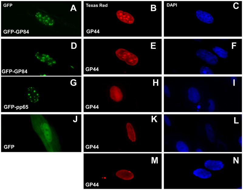
Figure 2. Co-localization studies of transiently expressed pGP84 and pGP44.
Transient plasmid expression assay of FLAG tagged pGP44 (pFLAGGP44 plasmid) in the presence of GFP tagged vectors: GFP tagged pGP84 (images A–F); GFP tagged pp65 (G–I); GFP control (J–L). Images M and N correspond to transient expression of pGP44 only cells. pGP44 detected by anti-FLAG primary antibody and secondary antibody anti-mouse IgG conjugated to TRITC. pGP44 images B, E, H and M. Cells counterstained with DAPI to identify cell nucleus (right column). Image A and D, pGP84 expressed as N-terminal tagged GFP protein (pGFPGP84). Image G, pGFPpp65 (HCMV pp65 N-terminal GFP tagged). Image J, control GFP plasmid pGFP-C1 (Clontech). Protein cellular localization determined at 12 hr post transfection as described in materials and methods.

