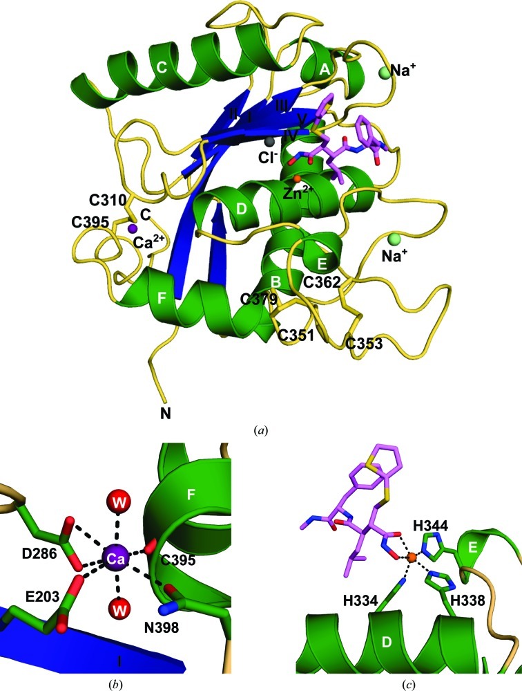
Figure 2.
Crystal structure of ADAM-8. (a) Ribbon diagram of the ADAM-8–batimastat complex. Molecule D was used for this diagram and therefore for most of the discussion. Secondary-structural elements are shown with α-helices in green (labeled A–F), β-strands in blue (labeled I–IV) and coils in wheat. The inhibitor batimastat is shown in lavender. Ions observed in this structure are represented as spheres. (b) Coordination of the Ca2+ ion (purple). Secondary-structural elements of ADAM-8 are displayed as in (a), with side chains shown in green. Two water molecules (red) complete the coordination sphere. (c) Coordination of the catalytic Zn2+ ion (orange). Histidine residues from the canonical zinc-binding motif are shown in green and batimastat is shown in lavender.

