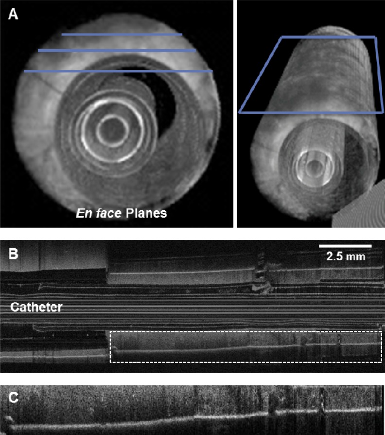
Fig. 5.
Visualization of longitudinal (long-axis) en face planes of tissue data prior to tissue biopsy. (A) Three-dimensional renderings of the acquired OCT data set, illustrating orientation of en face planes relative to catheter and needle. (B) En face plane extracted at a plane that includes not only the tissue but also the transparent plastic tube and sheath housing the OCT catheter. (C) Enlarged region of imaged tissue, from the region indicated by the white dashed box in (B).

