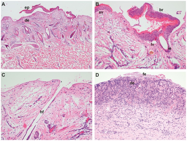
Figure 2. Histopathological features of DEB.
Biopsy of the ear of case 1: (A) Note that on the lateral borders of the biopsy the epidermis (ep) is missing and, where present, the epidermis is detached from the underlying dermis (de). The epidermis is cleanly separated from the dermis and the basal cells are intact. H&E, 25×. (B) Higher magnification: Note the intact basal cells of the blister roof (br), larger vacuoles (lv) and a small vesicle (sv) along the basement membrane zone and separation of the infundibular epithelium (ie) from the surrounding connective tissue. H&E, 200× (C) Note that the epidermal and infundibular epithelium of the hair follicle (hf) is missing and the surface is covered by homogenous proteinaceous material. H&E, 200×. Biopsy of the right hind leg of case 3: (D) Necrotic superficial dermis (de) characterized by fibrinous exsudation (fe), cellular debris, dense amounts of degenerate inflammatory cells, and hemorrhage. H&E, 100×.

