
Figure 3. SA2L contains NLS functional in yeast between 32K and 47K.
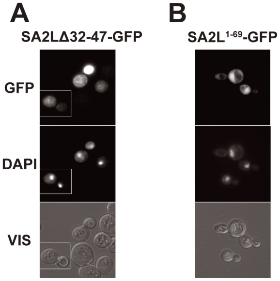
(A) – cells expressing fusion protein SA2LΔ32–47-GFP. (B) – cells expressing SA21–69-GFP. Compare Figure 1C. DNA was stained with DAPI. GFP represents fluorescence of fusion proteins, VIS – transmitted light. Column (A) shows a composite of two fields from a single experiment but photographed as separate images, as marked.