
Figure 5. SA1 contains NLS functional in yeast between 34K and 53K.
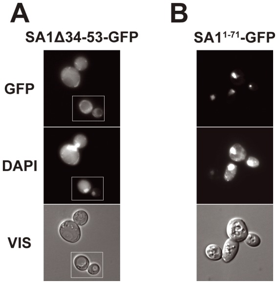
(A) – Cells expressing SA1Δ34–53-GFP. (B) – Cells expressing fusion protein SA11–71-GFP. DNA was stained with DAPI, GFP represents fluorescence of fusion proteins, VIS – transmitted light. Column (A) shows a composite of two fields from a single experiment but photographed as separate images, as marked. For subcellular localization of intact.