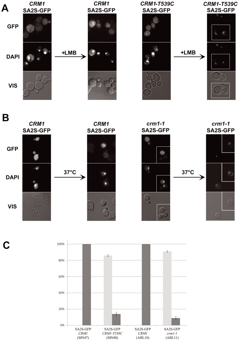
Figure 6. SA2S shuttles between nucleus and cytoplasm in yeast cells.
(A) Subcellular localization of SA2S-GFP was analyzed after addition of LMB (Crm1p inhibitor) to 40 ng/ml to cells in logarithmic phase of growth. Strain CRM1-T539C bears LMB-sensitive version of Crm1p. Fourth column shows a composite of two fields from a single experiment but photographed as separate images, as marked. (B) Localization of SA2S-GFP protein was analyzed in thermo-sensitive crm1-1 mutant. Transfer of cells grown at 30°C to 37°C for 30 minutes caused nuclear shift of the fusion protein in 100% of cells. Third and fourth columns show a composite of two fields from a single experiment, as marked. On the right in (A) and (B) control experiments in wild-type yeast are shown. DNA was stained with DAPI, GFP represents fluorescence of fusion proteins, VIS – transmitted light. (C) Frequencies of cells localized predominantly to the cytoplasm (black) or to the nucleus (gray) in strains bearing CRM1-T539C (LMB-sensitive) or crm1-1 (thermo-sensitive) versions of Crm1p, following LMB treatment or temperature shift, respectively. MN47 and ABL10 are corresponding control strains bearing wild type CRM1 gene, subjected to the same treatments.

