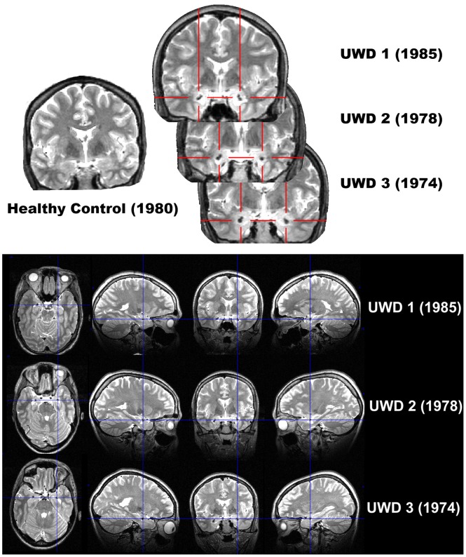
Figure 2. Structural MRI scans showing bilateral amygdala calcification in UWD.
Figure 2a: Coronal view T2-weighted MR-images of the three UWD subjects and one control subject with year of birth. Crosshairs indicate calcified brain damage. Figure 2b: T2-weighted MR-images in all three planes of the three UWD subjects. Crosshairs indicate the location of calcified brain damage bilaterally.

