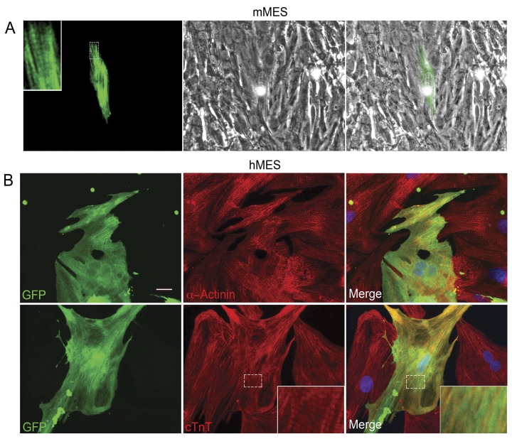
Figure 1. Bone marrow-derived MSCs in co-culture with cardiomyocytes.
(A) A nascent cardiomyocyte derived from mMSCs of a βMHC-YFP genotype mouse at 8 days in co-culture with cardiomyocytes. The βMHC-YFP cell is pseudo colored green for visualization of the striations. Left panel shows novel endogenous expression of βMHC-YFP fluorescence along stress fibers and striations (magnified in insert). Middle panel shows surrounding rat cardiomyocyte. Merged images in right panel. (B) GFP-hMSCs co-cultured with cardiomyocytes for 16 days and immunostained for α-actinin or troponin T ([23] and Supporting Information S1). Acquisition of a differentiated cardiac phenotype is demonstrated in the inserts where cardiomyocyte striations are visible. Nuclear DAPI in blue. Scale bar = 20 µm.

