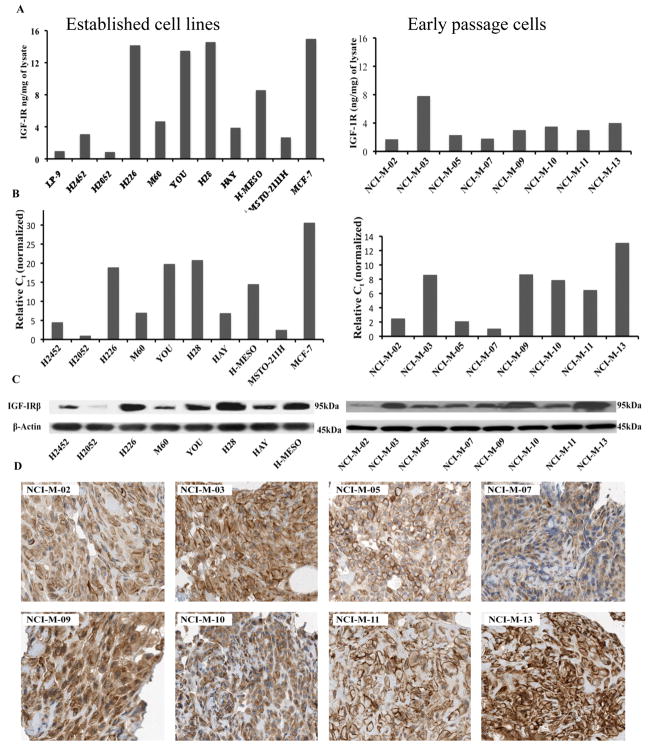
Figure 1.
Determination of IGF-IR in mesothelioma cells. (A) IGF-IR level was quantitated using an ECL-based IGF-IR sandwich immunoassay. Results are expressed in terms of ng/mg of cell lysate. The breast cancer cell line MCF-7 and normal mesothelial cell line LP-9 were used as a positive and negative controls respectively. (B) IGF-IR mRNA in mesothelioma cell lines was determined with real-time qPCR analysis. mRNA levels were normalized to GAPDH mRNA levels. (C) IGF-IR protein was detected by immunoblotting with an anti-IGF-IR mAb. Experiments were done in triplicate and representative image has been shown for both IGF-IR and the loading control β-actin. (D) Tissue sections obtained from paraffin embedded cell blocks of early passage mesothelioma cells were evaluated for IGF-1R expression by IHC using an anti-IGF-IR polyclonal antibody. All of these early passage cells show both membranous and cytoplasmic staining for IGF-IR.

