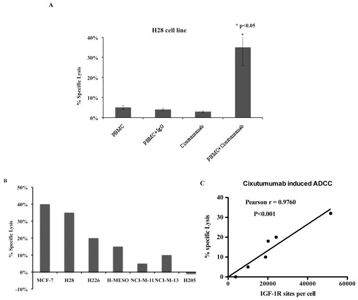
Figure 5.
Inductions of ADCC by cixutumumab in mesothelioma cell lines. Cells were incubated in the 96-well plate with 100 μg/mL of cixutumumab or a control human IgG at 37°C for 1 hr followed by addition of the effector human peripheral mononuclear cells. The percentage of specific lysis was calculated using lactate dehydrogenase assay in (A) H28 cell line and (B) in panel of mesothelioma cell lines with varying degrees of IGF-IR expression. (C) Pearson correlation analysis was performed using Graph pad to show the correlation between ADCC induced by cixutumumab and IGF-IR sites/cell expressed by each cell line.

