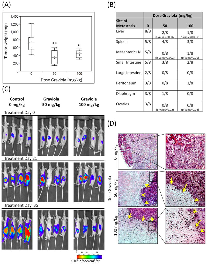
Figure 5. Evaluation of Graviola Extract in pancreatic cancer orthotopic xenograft model.
(A) Pancreatic tumor weight results after treatment with Graviola extract. CD18/HPAF-Luciferase cells were injected orthotopically in the pancreas of athymic nude mice. After 1 week of tumor growth, oral gavage treatment of PBS-suspended Graviola extract was given daily for 35 days (N=8). Data is presented as box plots of the mean tumor weight of mice in each treatment group. (*p-value = 0.006; **p-value=0.0008, compared to tumors of PBS-treated mice); (B) Major sites of metastasis in each treatment group. Results are presented as number of animals having metastasis out of total number of animals per group. Statistical analysis was done comparing Graviola extract-treated mice with untreated mice (0mg/kg Graviola extract); (C) In vivo biophotonic imaging of pancreatic tumors during the course of treatment with Graviola extract. Representative IVIS images of mice from different treatment groups are shown (D) Hematoxylin and Eosin (H&E) staining of paraffin embedded pancreatic tumors. Images on the right (20X) are magnified areas from the images located at the left (10X). Yellow arrows in H&E sections represent necrotic areas in tumors from mice treated with Graviola extract.

