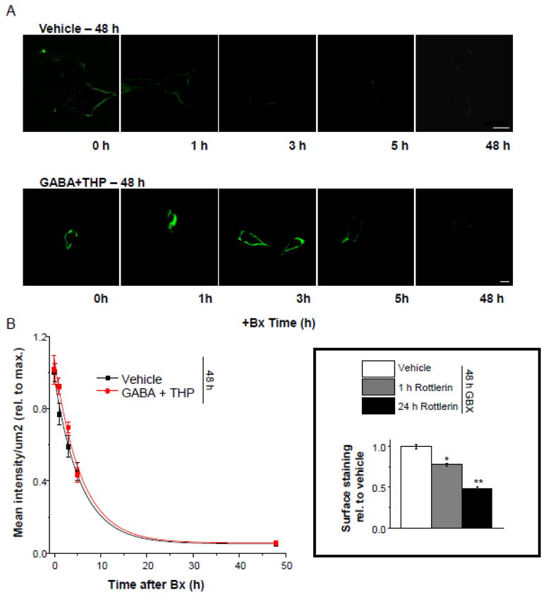
Figure 10. The rate of removal of surface α4Fβ2δ expression after 48 h gaboxadol treatment.
Transfected HEK-293 cells were treated with vehicle or gaboxadol (GBX, 10 μM) for 48 h. Where indicated, botulinum toxin b (Bx, 5 nM) was added to block receptor insertion across a time-course (1, 3, 5, or 48 h). A, Representative confocal images (63X) of α4 FLAG staining (488 λ, green) after treatment with Bx for the indicated time periods, administered alone (top) or in addition to 48 h GBX (bottom). scale bars, 10 μm. B, Averaged data. The rate of disappearance of α4F labeling is plotted as a function of time (n=5 cells per time point). The plot was best fit to a single exponential decay function, which was not altered by GBX treatment. Inset, Effects of rottlerin (10 μM), a PKC- δ inhibitor, on α4FLAG surface labeling after 48 h treatment with gaboxadol. ANOVA, F(2,9) = 8.3, P = 0.0091; *P<0.05 vs. Vehicle; **P<0.05 vs. 1 h GBX.

