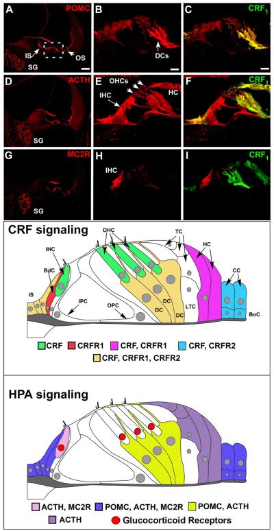
Figure 4. The cochlea expresses an HPA equivalent signaling system.
Immunofluorescent labeling of POMC, ACTH, and MC2R reveals expression of classic HPA components in the cochlea. (A, D, G) POMC and ACTH are expressed in inner and outer sulcus cells lining the cochlear duct (IS and OS, respectively). MC2 is expressed in these regions to a lesser extent (C). All components are expressed in the spiral ganglion cells (SG). The organ of Corti region is boxed in A and is the region from which the higher magnification illustrations were produced. (B) Higher magnification of the organ of Corti region reveals intense POMC labeling in the Deiter’s Cells (DCs) and this expression overlaps with CRFR1-GFP label (C). (E, F) ACTH shows less immunolabeling in Deiter’s cells compared to POMC, but an intense labeling of the inner hair cell (IHC). (H, I) Finally, in the organ of Corti region, MC2R shows an intense and specific labeling for the IHC and a lack of Deiter’s cell labeling. Bottom panels- Expression of CRF, CRFR1, and HPA components in the cochlea is mapped for clarity. CRF signaling molecules are expressed in the cochlea as defined in the immunostaining panels, and are re-created here in schematic fashion. Cells of the inner sulcus (IS) express CRF, CRFR1, and CRFR2. CRF alone is expressed in the inner and outer hair cells (IHC, OHC respectively) of the cochlea, shown in green. These CRF-positive sensory cells are juxtaposed by support cells such as the border cell (BdC, shown in red), which expresses CRFR1, and the Deiter’s cells (DC, cells directly below the outer hair cells and shown in dark yellow) that express CRF, CRFR1, and CRFR2. In addition to the Deiter’s cells, Tectal cells (TC) and Lateral Tunnel Cells (LTC) are directly apposed to the outer hair cells and Deiter’s cells, but do not express any CRF signaling components. Support cells located more laterally include the Hensen’s cells (HC), which flank the Tectal cells and Lateral Tunnel Cells laterally and express CRF, CRFR1, and the Claudius cells (CC) and Boettcher cells (BoC), which express CRF and CRFR2. Thus there is a potential for juxtacrine interaction between hair cells and support cells in their immediate vicinity. The inner sulcus cells medial to the border cell and support cells lateral to the organ of Corti express CRF, CRFR1, and CRFR2, suggesting autocrine and paracrine communications in these peripheral support cells that could also involve the hair cell populations. Finally, molecules of the classic HPA signaling system are expressed in the cochlea as defined in the immunostaining panels, and are schematically mapped for clarity. POMC, ACTH, and to a lesser extent, MC2R are expressed in inner sulcus cells, border cell near the IHC, and the lateral-most support cells which include Claudius cells and Boettcher cells (blue). Deiter’s cells express POMC and ACTH (depicted in yellow), but with little to no expression of MC2R. ACTH and its receptor, MC2R, are expressed in the IHC (pink), suggesting a convergence of HPA signaling on the afferent auditory transducer. ACTH alone seems to be expressed in the Hensen’s and Tectal cells (purple), with no discernable POMC expression found to date. Localization of previously described glucocorticoid receptors within the organ of Corti (based on (Terakado et al., 2011) is indicated by red nuclei, and demonstrates spatial proximity between cells expressing ACTH and MC2R and cells expressing glucocorticoid receptors. Scale bar in A represents 60μm and pertains to A, D, G. Scale bar in B represents 10μm and pertains to all other panels. HC = Hensen’s cell. CRFR1 designates CRFR1-GFP immunolabeling. (all figures reprinted from (Graham and Vetter, 2011) with permission. With permission, schematic figures were re-drawn and annotated from an original provided by Dr. M. Charles Liberman, Mass Eye and Ear Infirmary, Boston, MA)

