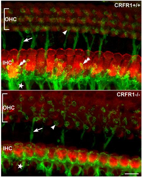
Figure 6. Elimination of CRFR1 leads to abnormal afferent fiber innervation to inner hair cells (IHC).
Afferent fibers and their most distal endings were visualized using antibodies directed against sodium-potassium ATPase α3 subunit (NKA, green). IHCs were immunolabeled using antibodies against myosin VI (red). Whole-mount sections of matched cochlear middle turns were imaged in CRFR1 wild type (top) and CRFR1 null (bottom) mice. Significant changes to hair cell width (modiolar to pillar along the y axis) are evident following loss of CRFR1 expression. In the CRFR1 null mice, afferent fibers (star) were found on all sides of the IHCs, and were decorated with immunoreactive puncta that outlined the termination of the fibers (double arrow in top panel). These fibers appeared stunted in CRFR1 null mice (bottom panel), with few endings reaching toward the pillar side of the inner hair cell, and none exhibiting the immunoreactive puncta observed in wild type mice. Additionally, moderate disorganization of the efferent fibers crossing the tunnel of Corti (arrows in top and bottom panels) and efferent innervation to the OHCs (single arrowhead) was also observed in the CRFR1 null mice. (reprinted from (Graham and Vetter, 2011) with permission)

