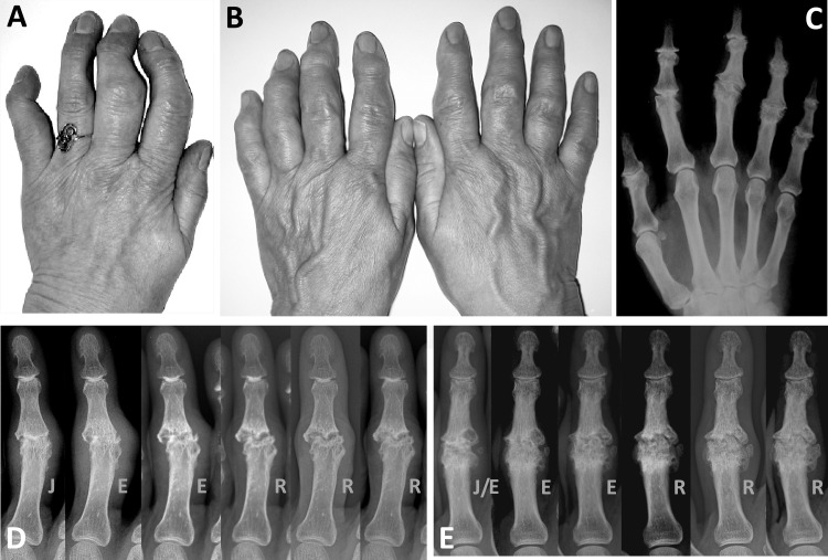
Figure 1.
Clinical appearance of erosive osteoarthritis of the interphalangeal finger joints is illustrated in (A) where erosive progression and subsequent remodelling caused functional inability of the second, third and fifth digit, and in (B) where almost all interphalangeal joints of both hands were affected and disabled. Remodelling of the interphalangeal joints in (B) after the erosive process is shown on the corresponding radiograph (C). Typically, metacarpophalangeal joints are spared in this condition. (D and E) Erosive progression and subsequent remodelling in two different proximal interphalangeal (PIP) joints is shown in a series of radiographic images taken at 6-month intervals. PIP joints in which the synovial joint space has disappeared (‘J’ phase) pass through the erosive ‘E’ phase and eventually remodel (‘R’).

