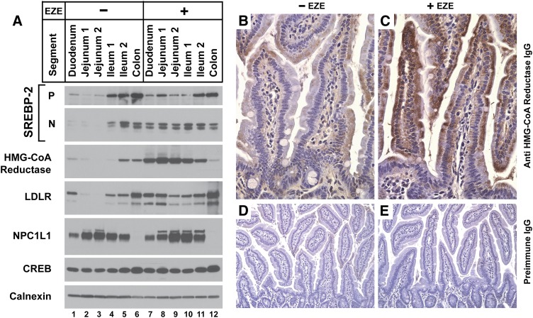
Fig. 2.
Anatomic distribution of ezetimibe's effect on SREBP-2 and its key effectors in the intestines. (A–E) Ages 12- to 13-week-old male C57BL/6J mice were fed chow diets with or without 0.01% ezetimibe in the diet for five days. (A) Immunoblot analysis of nuclear extracts and membrane fractions from enterocytes from sequential segments of intestine. Intestines were divided into five anatomic segments from duodenum to colon (see Materials and Methods) arranged in proximal to distal order. Enterocytes (four mice per group) were fractionated; 20 μg aliquots of the membrane and nuclear extract fractions were subjected to SDS-PAGE and immunoblot analysis. Immunoblots of CREB and calnexin were used as loading controls for the nuclear extract and membrane fractions, respectively. (B–E) Immunohistochemical analysis of HMG-CoA reductase in jejuna of untreated mice (B) or those who received ezetimibe (C), magnification 40×. Jejuna from untreated mice (D) and ezetimibe-treated mice (E), probed with preimmune antisera, magnification 20×.

