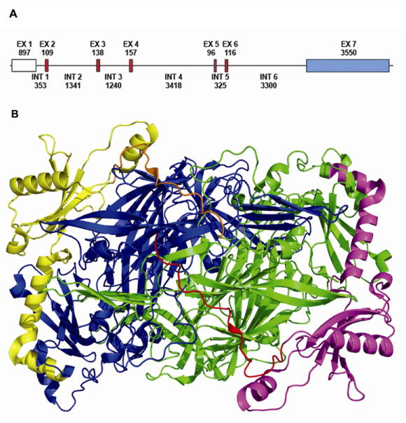
Figure 1. LOX gene and protein structure.
A, Exons are presented as boxes separated by introns (lines). The size of each exon and intron is shown in base pairs. The exons shaded in red encode amino acids sequences that are conserved in all lysyl oxidase family members. The exon shaded in blue contains the 3′ UTR sequence. B, 3D Crystal structure of Pichia pastoris LOX (Pymol). The molecule is presented as a dimmer with signal peptide domain (red and orange), pro-peptide (yellow and magenta) and C-terminal domain (green and blue) (Duff et al., 2003).

