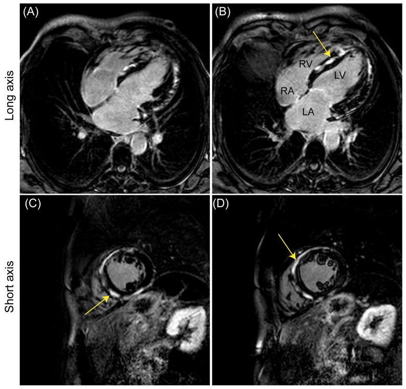
Figure 5.
Late gadolinium enhancement (LGE) long (A–B) and short (C–D) axis images acquired using a phase-sensitive inversion recovery (PSIR) sequence in a patient with suspected hypertrophic cardiomyopathy. An adaptive gating window with a user-defined efficiency of 40% was used for data acquisition resulting in an average navigator gating window of 2.6 mm. There were extensive, contiguous areas of focal hyper-enhancement (arrows) in the basal to mid antero- and infero septum, the basal to mid anterior and inferior segment, as well as the entire distal and apical walls.

