Figure 2.
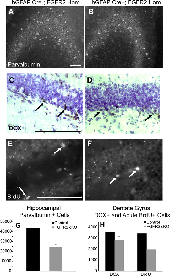
8–10 month old FGFR2cKO mice lacked cellular elements in hippocampus in adulthood. A,B: Immunocytochemistry for parvalbumin in CA showing decreased parvalbumin neuron density in FGFR2cKO as compared to control mice. C,D: Doublecortin (DCX) immunocytochemistry with cresyl violet counterstaining showing decreased DCX cell number in FGFR2cKO DG as compared to control mice. E, F: Immunocytochemistry for BrdU (1 hour after BrdU tracer injection) in control and FGFR2cKO DG. Arrows indicate DCX+ and BrdU+ cells. G: Histogram of average parvalbumin+ cell number in hippocampus of control and FGFR2cKO mice (n=5; 5). H: Histogram of average DCX+ and BrdU+ cell number in DG of control and FGFR2cKO mice (n=5; 6) (*= p<0.05). Scale bar= 200 µm.

