
FIGURE 5. Co-culture of thapsigargin-treated target cells with BMDC induces and enhances pro-inflammatory cytokine production in a calreticulin-independent manner.
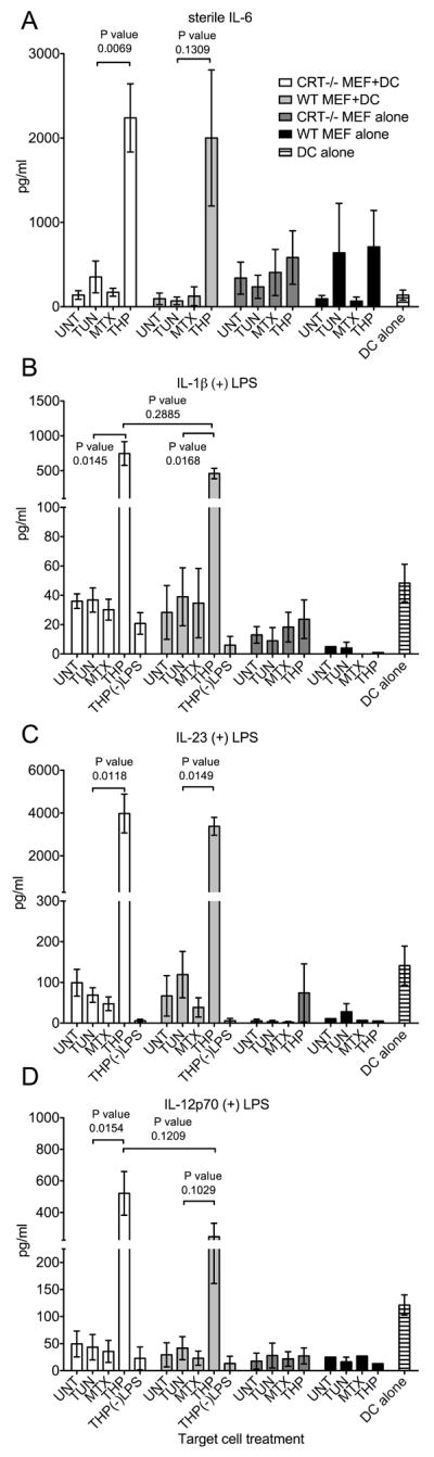
(A-D) Cytokine production by DCs, MEFs, and DC+MEF co-cultures. WT or CRT-/- MEF target cells were treated with TUN, MTX or THP for 5.5 or 6.5 hours or left untreated. Adherent target cells were then harvested, washed and incubated in media alone (MEF alone) or co-incubated with BMDC (MEF+DC) in the absence (A) or presence (B-D) of 0.5-2 ng/ml LPS for 18.5-23.5 hours and the concentrations of the indicated cytokine were measured in duplicate by ELISA. Wells of DC incubated in the presence (B-D) or absence (A) of LPS were included in each experiment as an additional control (DC alone). The THP(-)LPS bars indicate conditions where thapsigargin-treated WT MEF or CRT-/- MEFs were incubated with DC, but in the absence of LPS, to illustrate the LPS dependence of enhanced production of indicated cytokines. The data from co-incubations of DC and MEFs show the mean and standard error of 3 (WT) or 5 (CRT-/-) experiments. The data from MEFs alone or DC alone show the mean and standard error of 1-3 experiments in the presence of LPS, or 2-4 experiments in the absence of LPS. The p-values from two-tailed, paired t-tests are indicated.