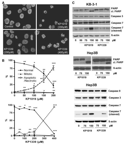
Fig. 2.
Comparison of the apoptosis-inducing potential of KP1019 with KP1339. a Induction of apoptosis in KB-3-1 cells was determined after treatment for 24 h. 4′,6-Diamidino-2-phenylindole (DAPI) staining of nuclei is shown in untreated controls and cells treated with the drug concentrations indicated. b Morphological features of 300–500 nuclei of at least two slides for each concentration were analyzed by DAPI staining. The percentages of normal, mitotic, and apoptotic/necrotic nuclei at the concentrations indicated are shown. c Apoptosis-induced cleavage of poly(ADP-ribosyl)polymerase (PARP), caspase 7, and caspase 3 in KB-3-1 and Hep3B cells after 24-h treatment was determined via western blot. The antibodies used are described in “Materials and methods”

